Surgeons at Och Spine at NewYork-Presbyterian are leading the field with years of historic collaboration between orthopedic surgery and neurosurgery to deliver high-quality pediatric spine care.
Dr. Michael Vitale spearheaded this approach for pediatric spine care more than two decades ago, which has delivered best-in-class outcomes for the most complex spine patients. Today, he collaborates on these cases with Dr. Taemin Oh.
The advantages of a joint orthopedic surgery and neurosurgery model include shorter surgical times, lower blood loss, and improved safety.
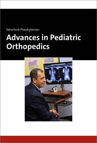
When it comes to pediatric spinal surgery, both neurological spine and orthopedic spine surgeons play critical roles. The specialties often collaborate on adult spine surgeries; however, only a few programs around the country have implemented a similar model in pediatrics. At Och Spine at NewYork-Presbyterian, pediatric orthopedic spine and neurological spine surgeons have spearheaded an integrated model of care that brings in both specialties as equal partners in the planning and delivery of pediatric spinal surgery.
Michael G. Vitale, MD, chief of pediatric spine surgery at NewYork-Presbyterian Morgan Stanley Children’s Hospital and Och Spine at NewYork-Presbyterian, and Taemin Oh, MD, a pediatric neurosurgeon and director of pediatric neurosurgical scoliosis surgery at Och Spine at NewYork-Presbyterian and Columbia, are setting an example on the critical ways the two specialties can collaborate on complex pediatric spine surgeries.
“For us to be at our best, we need to get out of the thinking about surgery as cut to close. The surgical episode starts way before that in terms of understanding the problem, the family, the social situation, the needs and priorities of the patient, and then planning accordingly.” — Dr. Michael Vitale
“Neurological spine surgeons are better at handling nerves, working around the spinal cord, and managing cerebral spinal fluid,” says Dr. Vitale. “Conversely, orthopedic spine surgeons have a deep understanding of the bones and biomechanics of the spine.”
The goal is to be “proactive as opposed to reactive,” Dr. Oh explains, so he and Dr. Vitale work together on a surgical plan, mapping out the workflow for the procedure, perform the surgery in tandem, and see patients together after surgery.
“For us to be at our best, we need to get out of the thinking about surgery as cut to close,” Dr. Vitale says. “The surgical episode starts way before that in terms of understanding the problem, the family, the social situation, the needs and priorities of the patient, and then planning accordingly.”
Tackling Complex Cases
Dr. Vitale and Dr. Oh recently completed a complex case together, successfully performing a hemivertebrectomy and posterior spinal fusion in an 11-year-old with progressive congenital scoliosis with a L5 hemivertebra. The hemivertebra, which caused 70 degrees of scoliosis, needed to be excised and the spine fused, so Dr. Vitale and Dr. Oh devised a surgical plan that involved Dr. Oh performing the hemivertebrectomy and Dr. Vitale performing the spinal fusion.
“This was a high-risk surgery because we had to remove that whole vertebrae from the spinal cord and the nerve roots,” says Dr. Vitale. “The nerve roots, especially at the bottom of the spine, are very sensitive to mobilization and there’s potential for postoperative weakness and bowel and bladder dysfunction. Working with a neurosurgeon like Dr. Oh allows me to utilize the expertise from both specialties to ensure that the patient has the best outcomes possible.”
“With the two specialties working together, we’re able to provide the absolute best for the patient – the surgery time is shorter, blood loss is lower, and it’s safer for the patient overall.” — Dr. Taemin Oh
The surgeons used intraoperative surgical navigation to visualize the bone and the nerve roots. Dr. Vitale placed screws above and below the hemivertebrae and Dr. Oh assisted him. A laminectomy was performed to remove the caudal aspect of L5a. Dr. Oh removed the hemivertebrae with Dr. Vitale assisting. They used drills and other tools while carefully retracting the nerve roots and protecting the spinal cord. Once the vertebrae was removed, rods were then measured, cut, and contoured and bent into coronal and sagittal alignment to correct the spinal deformity. The rods were inserted, and the screws were appropriately torqued down.